
Diagnostic imaging services in Henryetta, OK
Hillcrest Hospital Henryetta’s medical imaging center provides patients with a wide range of imaging tests to help detect and diagnose various conditions. Using advanced imaging technology and leading-edge techniques, our radiologists offer tests like bone density scans, computerized tomography (CT) scans, magnetic resonance imaging (MRI), and more. We pair expertise with compassion to ensure you receive the exceptional imaging services you and your loved ones deserve.
Imaging tests we provide
From angiography and X-rays to complex endoscopic procedures and digital mammography, we offer a full spectrum of advanced imaging services delivered by caring, knowledgeable technologists. We utilize leading-edge technology to provide the latest in diagnostic techniques.
Click below to explore our services in greater detail.
Bone density (DEXA) scan
This advanced X-ray technology measures bone loss and is especially effective in detecting osteoporosis. It provides detailed bone density assessments.
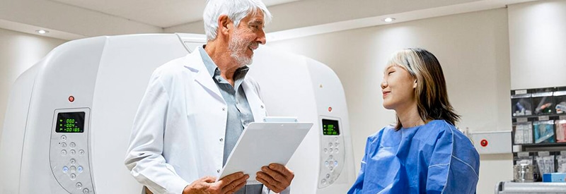
Computerized tomography (CT) scan
A CT or CAT scan combines X-ray and computer technology to show highly detailed, 3D images of any part of the body, including bones, muscles, fat, organs and blood vessels. Scans can also be performed using a contrast solution (either swallowed or injected) to make tissues and vessels more visible. We also provide low-dose lung CT screens.
Mammography
Mammography is an X-ray exam of the breasts used to screen for or diagnose breast cancer.
MRI
MRI scans are a diagnostic procedure that combines a powerful magnet, radio waves, and computer technology to provide detailed images of tissues, muscles, nerves and bones. Because MRIs use magnetic force and radio waves to create images, there is no radiation exposure during the procedure. MRIs are often used instead of CT to study soft tissues or organs because bones do not obscure the organs and soft tissues as they do with CT imaging.
MRI can be used to assess everything from ruptured discs in the spine to detecting brain tumors and vascular diseases through techniques such as diffusion scanning, or angiography, which evaluates blood flow and detects brain aneurysms or blood vessel abnormalities. Angiography is also used to visualize renal, carotid and vertebral arteries, or examine the aorta for aneurysm.
Ultrasound
Ultrasounds (or sonography) use reflected sound waves to create real-time images of soft tissues, including muscles, blood vessels and organs. Because sound waves are used, there is no radiation exposure during this procedure. Although most commonly used to examine the fetus during pregnancy, it is also an effective tool for monitoring blood flow using Doppler ultrasound technology.
Ultrasound can also be used to discover abnormalities in organs, and detect narrowed arteries, clotted veins, or growths such as tumors and cysts. We offer cardiac, vascular, abdomen, small parts, gynecologic and obstetric, breast, and thoracentesis ultrasounds.